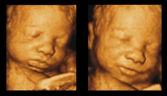
4D Ultrasound - Picture Your Baby's Face
 A typical ultrasound used to be a grainy, black and white image with the shadowy form of your baby. Fortunately, that's changed. Cabell Huntington Hospital uses 4D ultrasound to capture images of your unborn baby. Four-dimensional, or 4D, ultrasound takes three-dimensional ultrasound images and adds the element of time to the process. The result is live action images of your unborn child with far more detail and definition than any other imaging process available. A 4D ultrasound provides a sharper picture of your baby, and it serves as a powerful tool that can help physicians studying the baby's motion and behavior, the baby's surface anatomy and problems related to the mother's uterus and ovaries.
How can I get a 4D ultrasound?
 At the Perinatal Center, we attempt 3D/4D pictures when you are between 28-32 weeks into your pregnancy because the best pictures can generally be obtained in the early weeks of the 3rd trimester. If your doctor determines that an ultrasound is required during that time, an order for the specific ultrasound test will be sent to our office and we will attempt a 3D/4D image during that specific ultrasound scan. It is important to know that while we will do our best to get a picture like the one shown here, there are some uncontrollable factors that may keep us from obtaining that perfect 3D/4D image or picture. Some of the more common reasons we encounter are the location of the placenta, the position or size of the baby, low amount of amniotic fluid and maternal body shape and size.
Since an ultrasound order for a specific test is required from your physician (not just a request for 3D/4D pictures), your physician’s office staff will make arrangements for the appointment. Please do not hesitate to call the Perinatal Center staff with questions, as we will be happy to answer them at any time.
